
Peri-Implant Mucositis vs. Peri-Implantitis: Key Differences, Symptoms, and Treatment
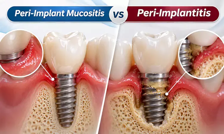
A dental implant can look and feel completely natural, but the gum tissue surrounding it still needs regular care. Redness, swelling, or bleeding around an implant may indicate inflammation, but these signs do not always mean that the supporting bone is affected. Understanding Peri-Implant Mucositis vs. Peri-Implantitis is important because the two conditions involve different levels of damage and may require different treatment approaches.
Peri-implant mucositis affects the soft tissue around an implant, while peri-implantitis also involves progressive loss of the supporting bone. This guide explains how to recognize the possible signs, how dentists distinguish between the conditions, and what treatment and preventive measures may help protect your implant.
Peri-Implant Mucositis vs. Peri-Implantitis: What Is the Main Difference?
The main difference between these two conditions is whether inflammation is limited to the soft tissue or has also affected the bone supporting the implant. Both are inflammatory diseases associated primarily with bacterial plaque accumulation around dental implants. However, their clinical features and potential consequences are not the same. In Peri-Implant Mucositis vs. Peri-Implantitis, the presence or absence of additional supporting bone loss is the most important distinction. Peri-implant mucositis can often be reversed with appropriate professional care and improved oral hygiene, whereas peri-implantitis requires more comprehensive assessment and may need surgical treatment.
What is peri-implant mucositis?
Peri-implant mucositis is inflammation of the soft tissue surrounding a dental implant without additional bone loss beyond the normal changes associated with initial healing. It is commonly linked to the accumulation of bacterial plaque around the implant and its restoration.
Common signs may include:
Redness or swelling of the gum around the implant
Bleeding when brushing or during gentle professional examination
Gum tenderness or irritation
Occasionally, suppuration around the implant
Although these symptoms may seem minor, they should not be ignored. Early professional treatment can help control inflammation before it develops into a more serious peri-implant condition.
What is peri-implantitis?
Peri-implantitis is an inflammatory condition that affects both the soft tissue and the supporting bone around a dental implant. In addition to signs of inflammation, the condition involves progressive bone loss that can compromise the support of the implant.
Possible clinical findings include:
Bleeding when the tissue around the implant is gently examined
Swelling, redness, or suppuration
Increased probing depth compared with previous examinations
Gum recession around the implant
Radiographic evidence of supporting bone loss
In more advanced cases, the loss of supporting bone may affect implant stability. However, not every patient experiences noticeable symptoms in the early stages, which makes regular dental examinations important.
Peri-implant mucositis vs. peri-implantitis: A comparison table
Comparison point | Peri-implant Mucositis | Peri-implantitis |
Affected tissue | Soft tissue around the implant | Soft tissue and supporting bone |
Main problem | Inflammation of the gum tissue | Inflammation accompanied by progressive bone loss |
Bleeding | May occur during brushing or gentle probing | May occur during gentle probing |
Swelling and redness | Common clinical signs | May also be present |
Bone loss | No additional pathological bone loss | Progressive supporting bone loss is present |
Diagnosis | Clinical examination and assessment of bone levels | Clinical examination, probing measurements, and radiographs |
Treatment | Professional plaque removal and improved oral hygiene | Non-surgical treatment and, when needed, surgical procedures |
Reversibility | Often reversible with appropriate treatment | Existing bone loss cannot be assumed to reverse; treatment aims to control disease and manage the defect |
The table highlights why symptoms alone cannot establish which condition is present. A dentist needs to examine the surrounding tissue and evaluate the bone levels before making a diagnosis.
If you are considering implant treatment or need advice about an existing implant, you can explore available Dental treatment georgia and Dental treatment tbilisi options.

What Are the Symptoms and Risk Factors of Peri-Implant Diseases?
Recognizing the signs of inflammation around a dental implant is an important part of protecting its long-term health. Both conditions can cause redness, swelling, and bleeding, so patients may not be able to distinguish between them at home. The comparison of Peri-Implant Mucositis vs. Peri-Implantitis becomes clearer when a dentist evaluates changes in gum tissue, probing measurements, and supporting bone levels. Some patients may experience discomfort or notice bleeding, while others may have few obvious symptoms. For this reason, the absence of pain does not necessarily mean that the tissues around an implant are healthy.
Here you can read about How to Compare Dental Clinics Abroad
Which symptoms should you look out for?
Contact a dental professional if you notice any of the following changes around your implant:
Bleeding during brushing or flossing
Persistent redness or swelling around the implant
Gum tenderness or discomfort
Pus or discharge near the implant
Changes in the appearance of the gum line
A feeling that the implant-supported restoration has changed position or become loose
A loose crown and a loose implant are not necessarily the same problem. A restoration may become loose because of a prosthetic or mechanical issue, while implant mobility can indicate a more serious problem with implant support. Both situations require professional evaluation rather than self-diagnosis.
What increases the risk of peri-implant mucositis and peri-implantitis?
Several factors can contribute to inflammation or increase the risk of disease around dental implants. The most important factor is the accumulation of bacterial plaque, but individual health conditions and the design or position of the restoration may also influence the outcome.
Relevant risk factors include:
Poor plaque control: Inadequate cleaning allows bacterial biofilm to accumulate around the implant.
Previous periodontal disease: A history of periodontitis is associated with a higher risk of peri-implant disease.
Smoking: Tobacco use is an important risk factor for complications around dental implants.
Uncontrolled diabetes: Poorly controlled blood glucose may increase the risk of inflammatory complications.
Difficult-to-clean restorations: The shape or position of an implant-supported crown may make effective cleaning more challenging.
Other patient and site-related factors: Implant positioning, prosthetic design, and individual soft-tissue characteristics may affect the ability to maintain peri-implant health.
Having one or more risk factors does not mean that peri-implantitis will definitely develop. It means that preventive care and professional monitoring may need particular attention.
How Do Dentists Diagnose Peri-Implant Mucositis and Peri-Implantitis?
A reliable diagnosis requires more than checking whether the gum is red or bleeding. When assessing Peri-Implant Mucositis vs. Peri-Implantitis, dentists examine the soft tissue around the implant and look for signs that the supporting bone has changed. They also consider the patient's previous dental records, including earlier probing measurements and radiographs. Comparing current findings with these baseline records can help identify changes that may not be obvious during a routine visual examination. The diagnostic process is therefore based on a combination of clinical findings and imaging rather than a single symptom.
Clinical examination and periodontal probing
During an examination, the dentist may gently assess the tissue around the implant using a periodontal probe. This helps measure the depth of the space between the gum and the implant and identify bleeding or suppuration.
The results are particularly useful when compared with measurements taken after the implant-supported restoration was placed. An increase in probing depth can have different explanations, including inflammation-related swelling, so it should not be interpreted on its own as proof of peri-implantitis.
Dental X-rays and supporting bone assessment
Dental radiographs allow the dentist to evaluate the bone levels around the implant. Comparing current images with earlier baseline radiographs helps determine whether additional bone loss has occurred.
When previous examination data are unavailable, internationally accepted case definitions use a combination of findings to support a diagnosis of peri-implantitis. These include bleeding or suppuration on gentle probing, probing depths of at least 6 mm, and bone levels at least 3 mm apical to the most coronal portion of the intraosseous implant. These are professional diagnostic criteria, not measurements patients can use to diagnose themselves.
An accurate assessment also helps the dentist identify possible contributing factors, such as plaque-retentive restoration contours or retained dental cement around the implant-supported crown.
How Are Peri-Implant Mucositis and Peri-Implantitis Treated?
Treatment depends on the diagnosis, the extent of inflammation, the amount of supporting bone loss, and the patient's overall oral health. In Peri-Implant Mucositis vs. Peri-Implantitis, the treatment goal for mucositis is to control inflammation and restore healthy soft tissue conditions. Peri-implantitis requires a more detailed treatment plan because the supporting bone is also affected. In both situations, controlling bacterial plaque and addressing contributing risk factors are important parts of care. The dentist or periodontist determines the appropriate approach after evaluating the implant and surrounding tissues.
Treatment options for peri-implant mucositis
The usual first step is professional mechanical plaque removal combined with improved daily oral hygiene. The dentist cleans the implant surface and restoration using suitable instruments and explains how the patient can maintain the area at home.
Treatment may involve:
Professional removal of plaque and deposits around the implant
Cleaning the accessible surfaces of the implant-supported restoration
Instructions on using suitable interdental brushes or implant-specific floss
Assessment of the restoration shape to make daily cleaning easier
A follow-up appointment to check whether inflammation has improved
Routine local or systemic antibiotics are not recommended as standard treatment for uncomplicated peri-implant mucositis. Additional measures should be considered by the treating professional only when clinically indicated.
Treatment options for peri-implantitis
Peri-implantitis treatment is more complex because the dentist must manage both inflammation and the bone defect. Treatment usually begins with a non-surgical phase, including oral hygiene instruction, risk-factor control, professional cleaning, and assessment of the implant-supported restoration.
Depending on the response and severity of the condition, further treatment may be considered:
Non-surgical debridement: Removal of bacterial deposits from accessible implant and restoration surfaces.
Correction of contributing factors: Assessment of restoration contours, retained cement, and other factors that make plaque removal difficult.
Surgical treatment: In selected cases, surgery may be considered to access the affected area and manage the bone defect.
Regenerative or resective procedures: Depending on the shape and severity of the defect, a specialist may consider procedures intended to reconstruct or reshape the affected area.
Not every patient needs surgery. The treatment decision depends on the clinical findings and the response to initial therapy. Existing bone loss should not be assumed to regenerate fully, and outcomes vary according to the individual case.
How Can You Prevent Peri-Implant Diseases and Protect Your Dental Implants?
Prevention is essential for maintaining healthy gums and supporting tissues around dental implants. The most effective approach combines consistent home care with regular professional examinations. Understanding Peri-Implant Mucositis vs. Peri-Implantitis also helps patients recognize why early attention to gum inflammation matters: mucositis can often be managed before supporting bone becomes involved. Patients with a history of periodontal disease, smoking habits, or other risk factors may need closer monitoring. A personalized maintenance schedule helps the dental team identify changes and respond before they become more difficult to manage.
Daily habits that help protect your implant
To reduce plaque accumulation and maintain healthy peri-implant tissues:
Brush your teeth twice daily with a suitable toothbrush, paying attention to the gum line around the implant.
Clean between your teeth and around implant-supported restorations using interdental brushes or other tools recommended by your dentist.
Avoid smoking and seek professional support if you need help quitting.
Follow your dentist's advice for managing diabetes or other health conditions that may affect oral health.
Attend regular maintenance appointments so that plaque deposits and early signs of inflammation can be identified.
Contact your dentist if you notice bleeding, swelling, or other changes around the implant rather than waiting for your next routine visit.
Here you can read more about What to eat after dental implant surgery?
Why are regular dental check-ups important?
A dental implant requires ongoing care even after the final crown has been placed. During maintenance visits, the dental professional can assess the soft tissue, check probing measurements, remove deposits, and evaluate the restoration. Radiographs may also be recommended when needed to monitor bone levels. The frequency of these visits should be based on the patient's oral health, risk factors, and clinical findings rather than assuming that the same schedule is appropriate for everyone.
Patients researching Dental implant georgia or Dental implant tbilisi can also review the implant treatment information available through Alo.dr and discuss their individual needs with a dental professional.
How Should You Understand the Difference Between Peri-Implant Mucositis and Peri-Implantitis?
The key distinction in Peri-Implant Mucositis vs. Peri-Implantitis is whether the inflammation is limited to the gum tissue or is accompanied by progressive loss of the bone supporting the implant. Peri-implant mucositis can often be controlled with professional plaque removal and improved oral hygiene, while peri-implantitis may require more advanced treatment based on the extent of bone damage. Because both conditions can cause similar symptoms, bleeding or swelling alone cannot confirm the diagnosis. Regular dental examinations, appropriate imaging, and consistent home care are essential for protecting implant health over time.
Frequently Asked Questions About Peri-Implant Mucositis and Peri-Implantitis
1. Can peri-implant mucositis turn into peri-implantitis?
Yes. If inflammation remains uncontrolled, peri-implant mucositis may progress to peri-implantitis, which involves supporting bone loss. Early diagnosis and appropriate treatment can help reduce this risk.
2. Is peri-implantitis painful?
Not necessarily. Some people may have bleeding, swelling, or discomfort, while others experience few noticeable symptoms. A dental examination is necessary to assess the condition.
3. Can a dental implant be saved if peri-implantitis develops?
In some cases, treatment can control the disease and help retain the implant. The outcome depends on factors such as the amount of bone loss, implant condition, and response to treatment. A dentist or periodontist must assess each case individually.


Comments